Answers from Tartu Science Night 2023
One way that we link genetics to a neurodegenerative disease is when we observe that a particular disease runs in families. For example, in 1872, George Huntington, a 22-year-old physician in the US, was the first to officially describe a type of chorea (uncontrolled, jerky movements) in a disease that came to bear his name – “Huntington’s disease”. Dr Huntington was part of a long line of physicians, and by carefully reviewing his father’s notes, he was able to see that the condition was hereditary. See more on Huntington’s disease here.
Nowadays, we can look at the DNA of patients to link genetics to a disease. For example, in 2003, researchers examined DNA samples from a large family with many members who had autosomal dominant Parkinson’s disease. When a disease is autosomal dominant, it affects everyone who carries the mutation, and the disease does not skip generations. The researchers found that a single mutation caused increased expression of a protein called alpha synuclein and this increased expression of alpha synuclein caused their disease. Learn more about Parkinson’s disease here.
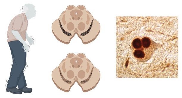
Left: A patient with Parkinson’s disease walks more slowly and has tremor (shakiness). Middle top: This image shows the area of the brain where the neurons that help control movement are found. In the middle bottom image, you can see that some of these neurons are lost – that is what happens in Parkinson’s disease. Right: In a patient with Parkinson’s disease, these neurons develop clumps of alpha synuclein called Lewy bodies. This image is adapted from Lancet Neurology 2015 Oct;14(10):1054-64. The cartoons were developed using BioRender.
With regard to how genetics and environmental factors interact, individuals carrying a particular mutation that causes Parkinson’s disease who live in the United States show a different disease duration when compared to their relatives who live in Italy – researchers believe environmental factors are the cause. In another example, following the Gulf war, many soldiers became ill with Gulf war syndrome, which causes problems with thinking, in addition to a host of other symptoms. The condition was due to exposure to a toxin, but only people with a mutation in a gene called PON1 developed the condition. The mutation in the gene lead to the expression of a defective Pon1 protein, that was less capable of breaking the toxin down. Thus, genetics and environmental factors do interact, and in fact, we believe that many diseases, including Parkinson’s disease and Alzheimer’s disease are caused by a combination of gene mutations and environmental factors.
When we use mice to “model” a particular disease, we must ensure that the cause of the disease in mouse and human is similar, and the symptoms displayed by the mouse is similar to the symptoms shown by patients. For example, excessive stress places humans at increased risk of depression, and stressed mice and rats also show symptoms of depression. A naturally occurring mutation in some mice leads to seizures and this mutation also leads to seizures in humans. In other lines of mice, we insert a mutant form of a gene that causes Alzheimer’s disease in humans into the mouse genome. All of these “models” are critical for greater understanding of depression, epilepsy and Alzheimer’s disease.
Mice are used all over the world to help understand human diseases. For example, in Parkinson’s disease, we know that the disease begins decades before diagnosis and this stage is very difficult to examine in humans because the individual is not ill. We can use mice and other animal “models” to understand disease mechanisms during these very early stages. We can also learn things like the best dose range to use for a particular therapeutic, or whether a sufficient amount of a therapeutic can get into brain. Therefore, through careful, ethical research using mice, we can learn a lot about human diseases and how to treat them.
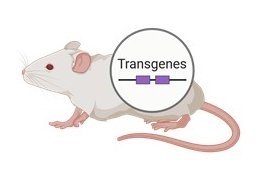
Transgenic mice are mice that carry a “transgene” within their DNA. For example, this could be a gene that expresses a mutant form of alpha synuclein that causes Parkinson’s disease in humans. Scientists can then study how this transgene affects the mouse, to better understand mechanisms underlying Parkinson’s disease. cartoons were developed using BioRender.
Learn more about the responsible and ethical use of animals in research here.
Learn why we need to do careful research in animals here.
Learn how our standards of care for our mice and the research we do with them is governed by the law in Europe.
Physical and emotional pain are indeed related – for example, depression is a serious disorder that causes intense emotional suffering in the patient. An individual who is depressed is more likely develop chronic pain conditions, but also, someone with chronic pain is more likely to develop depression.
In a further link, drugs used to treat depression modulate levels of the brain chemicals noradrenaline and serotonin – and the same drugs can be used to treat chronic pain.
Moreover, the drug Ketamine is used to control severe pain and can be used as a general anaesthetic, but new data show that it is also useful for some patients with depression.
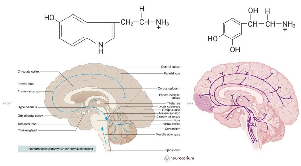
Top left: This molecule is called “Noradrenaline”. It is released from neurons in the brain and one of its functions is to help control emotion. Neurons that release noradrenaline are shown in the diagram on the bottom left (this image is from Neurotorium. You can see that their body is present in the base of the brain (see the blue dots of the “Locus coeruleus” for example). On the top right, you can see the molecule “Serotonin”. This is another molecule that is released from neurons in brain. Serotonin is critical for the control of our emotions. The bodies of these neurons are also found in the base of the brain and they send projections all over the brain, just like the noradrenergic neurons. Molecules and image on bottom right created using BioRender.
In rare cases, severe grief, anger or surprise can lead to physical pain like angina – this is called Broken Heart syndrome. It is most often seen in women, and it is caused by stress-related increased activity in an area of the brain called the amygdala. The amygdala controls emotions and some forms of learning and memory. Learn more about Broken Heart syndrome here.
Note that while under general anaesthesia, our brain is not capable of responding to pain or to any external signal. This is very important because the failure to respond normally to the pain of making an incision, for example, allows the surgeon to do their work.
All of this indicates that pain and depression share some brain pathways but we don’t yet fully understand why or how. Learn more about depression here.
Forgetting where you put the car keys or forgetting a monthly bill are examples of normal age-related memory loss. These problems don’t affect your ability to live your life and although annoying, they are part of our aging process.
More serious forms of memory loss can lead to problems like not remembering how to make a cup of tea, not remembering how to find your way home, not being able to figure out the correct date or having difficulty speaking with others. These may be signs of dementia, which is not normal aging. This kind of memory loss affects your ability to live your daily life.
We know that people who have higher childhood education levels and higher lifelong levels of education are less likely to develop Alzheimer’s disease, which is the most common form of dementia.
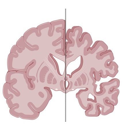
In this cartoon (created using BioRender), we can see a normal brain on the left and a brain affected by Alzheimer’s disease on the right. We can see “atrophy” or shrinking in the brain with Alzheimer’s disease, which is caused by the loss of neurons.
However, we must remember that our ability to go to college is affected by our socioeconomic status (our economic and social position relative to others).
A high socioeconomic status means that we can more easily go to college and it also means we eat better food, we are healthier and we have a better quality of life. So, education is not the only factor that protects us! As another example, people who work in jobs that require interaction with others are also less likely to develop Alzheimer’s disease.
As the saying goes – for your brain, we must use it or lose it!
Have a good diet and exercise regularly. Meet up with friends and have fun. Protect your head – wear a helmet when cycling and wear a seat belt in the car. All of these things, especially eating a proper diet and regular exercise, will help protect against memory loss.
Learn more about Alzheimer’s disease here.
Genetics account for about 50% of our intelligence.
One gene that has been studied is catechol-O-methyl-transferase (COMT). This gene encodes a protein that normally degrades dopamine in brain. This gene has some functional polymorphisms, which means that it has variants that differ by a single base position in its DNA. Individuals carrying some modified forms of the COMT gene show better working memory (short-term memory).
However, there are likely many genes that affect intelligence, and variations in each are likely to have a small effect.
In fact, super-intelligent people with intelligence quotients (IQs) of over 170 (the average IQ is 100), actually lack the many mutations that the rest of us have! Learn more about DNA here.
Also, remember that although 50% of our intelligence is controlled by our genes, 50% is controlled by something else – our childhood environment, our lifestyle, our diet, our healthcare, our education…
Also, although IQ is constant, we can improve our memory by regular training – this includes mental stimulation, for example education, and social interactions, as well as diet and exercise.
Sources:
- Mol Psychiatry. 2011 Oct; 16(10): 996–1005
- Biological Psychiatry. 2008 15 July, 137–144
A great place to start is coming to Brain Awareness Week at the University of Tartu! We are so happy you came!
Indeed, a special goal of ours was to make a space for you, our visitor, to talk about neuroscience. Another goal is to create a special webpage within ut.ee that is dedicated to neuroscience.
Some great resources for further information are within the Federation of European Neurosciences website, here.
The Society for Neuroscience is an American organisation and they have developed a great resource for people interested in the brain here.
If you would like to get involved in neuroscience, simply review the laboratories within University of Tartu and send the laboratory head an email! It is really that simple! The University of Tartu has many programs to help you get involved in research, and Talendid Tartusse is a great program to help highschool students see and do research. Learn more here.
I was standing about 2 metres away from a 1.5T MRI and within 15 minutes, I started to get a heavy feeling in my heart and I also started to lose my balance. After 30 minutes, I could feel arrhythmias. I am a young medical student and I do not have a pacemaker. The pain in my chest lasted for about 4 hours after leaving the room. These symptoms have now recurred three times in the same situation.
A thought-provoking question. Magnetic resonance imaging is considered to be very safe.
However, magnetic resonance imaging (MRI) depends upon a very strong magnet and so there should be no metal objects in the room that could dislodge and move, which could result in injury.
Also, the patient should have no implanted devices, such as metal stents, as they could dislodge.
Some heating (approximately 1°C) of your body surfaces can also occur, which is easily dealt with by your blood vessels, which expand slightly to dissipate the heat.
The United States Food and Drug Administration (FDA) has stated that magnets of up to 8 Tesla for adults and up to 4 Tesla for babies up to 1 month of age are safe. Most hospital MRI scanners are approximately 1-2 Tesla. An Israeli study of children whose mothers had been scanned while pregnant showed no harmful effects on these children. A study in Sweden of MRI safety revealed some issues with projectiles, where items had become dislodged by the powerful magnet, but no cardiovascular effects like those in this question.
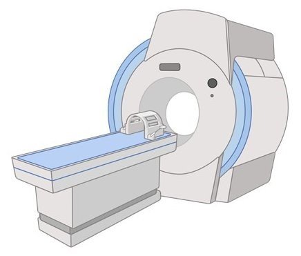
A magnetic resonance imager. Using powerful magnets, they help physicians see inside the body and in particular, magnetic resonance imaging helps create detailed images of “soft” tissue, e.g., brain. Cartoon created using BioRender.
However, magnetic resonance imaging is known to stimulate peripheral nerves. These are the nerves that help us feel touch, cold, heat, pressure and pain. Patients typically report tingling or vibration, hence, it is not considered to be dangerous. It is thought to be due to the changing magnetic gradients within the scanner, which then cause these neurons to fire, resulting in the tingling sensation. In animal models exposed to very high levels of changing magnetic gradients, which are not used in humans, some arrhythmias can occur. See more on this in the final link below. It is possible that you are much more sensitive to the gradients. If you need to have imaging done, please discuss your symptoms with your physician. Thank you for sharing this very interesting story!
Sources:
Another very interesting question. Osteoporosis is a condition that reduces bone strength or density and it can lead to fractures if left untreated.
Sometimes, certain populations do show an increased prevalence of specific diseases. In general, Caucasian women and men have a higher risk of osteoporosis, compared with, for example Asian people.
Approximately 1 in 5 women of 50 years of age or more have osteoporosis whereas about 1 in 20 men of the same age have osteoporosis. This is because of changes in hormones that occur at menopause – oestrogen levels decline. As oestrogen is very important for bone health, this loss in oestrogen places women at greater risk of osteoporosis.
Some other factors that can increase the risk of osteoporosis include lack of exercise, smoking, if your body cannot absorb vitamins properly, low weight, excessive alcohol, a family history of osteoporosis, poor diet, air pollution, and several diseases, such as diabetes, and some drugs, such as corticosteroids, a type of hormone that is sometimes used to treat conditions like severe asthma or Crohn’s disease.
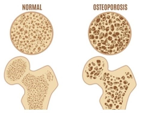
On the left, you can see what normal bone looks like. On the right, the beautiful honeycomb structure is weakened and the bone itself is less strong, which can lead to fractures. Image from here.
To lower your risk of osteoporosis, make sure that you eat well and take regular exercise. Calcium and vitamin D are important for bone health, so if you live in a country with poor light levels in winter, for example, talk with your physician about supplements. Osteoporosis is a “silent” disease so make sure to attend screenings because they can help identify reduced bone density before any problems, e.g., a fracture, occurs. With early diagnosis and proper treatment, patients can expect to live normally.
Source:
- Ther Clin Risk Manag. 2018; 14: 2029–2049
Great question. Unfortunately, cancer is the second leading cause of death worldwide after cardiovascular disease, but survival rates are improving due to better and earlier detection, better treatments and greater awareness among the general population.
For example, breast cancer is the most common cancer in women, but the number of breast cancer survivors in Europe has improved over the past 30 years. For men, the second-most common cancer type is prostate cancer, and if detected early, almost 100% of patients survive. Lung cancer is the most common cancer in men, and it also shows improved survival over the past 30 years. Countries that encourage control of smoking control have even better survival rates. However, smoking among women, and lung cancer in women, remains an issue in many countries.
The European Code Against Cancer shows several actions that EU citizens can take to reduce their risk of cancer. Some of these actions include not using any form of tobacco, reducing excessive sun exposure, maintaining a health body weight and exercising regularly. Also, make sure to eat well with lots of fruit and vegetables. Avoid excessive amounts of red meat and processed foods. Citizens should attend screenings and take part in vaccination programs where appropriate, for example, for Hepatitis B. View more here.
Sources:
- EClinicalMedicine. 2021 Jul 7;38:100985
- J Epidemiol Glob Health. 2023 Sep; 13(3): 407–421
- Lancet Respir Med. 2021 Sep;9(9):1030-1049
Fascinating question. Some people can, literally, remember everything that has happened to them. This is an extremely rare condition and is called hyperthymesia. Less than 100 people have been diagnosed with hyperthymesia.
For most of us, our memories are formed due to a collaboration of different areas of our brain – we remember smells, colours, feelings, together with facts and places, but our memories change with our experiences and so accuracy can change over time.
Some individuals with hyperthymesia say that it is a blessing, but others say it can be quite burdensome, as anything can trigger a flood of memories. Rather than forgetting their past choices and mistakes, they remember them. People with hyperthymesia can show some signs of obsessive-compulsive disorder and tend to categorise their memories and have some changes in brain structure, but it is not clear if this is the reason underlying their incredible memory, or that they change in their brain is a result of the many memories.
So, is being able to remember everything a benefit or a drawback? Well, that’s for you to decide. Would you like to remember everything? Would you like to remember some things and forget other things?
Sources:
Very interesting question. The vast majority of people can tell up from down, but actually, many people have difficulty in telling left from right – maybe up to 1 in 6 people.
We call this right-left confusion and it tends to be more common in women and left-handed people. This may be due to brain lateralisation. For example, men are typically more spatially aware (right hemisphere-related task), whereas women typically show more verbal ability (left hemisphere-related task).
This issue is of concern in medicine: wrong-sided surgery is a rare, but it is a recognised issue. A survey of male and female medical students in the UK showed that males were better at distinguishing right from left and that students who wished to become a surgeon were better than students who wished to become a general practitioner. But, right-handed people were as good as left-handed people.
For individuals who have issues telling left from right, there are strategies that can help. For example, look at your writing hand, or form an L shape with the thumb and forefinger. Or, think about the side of the road you drive on. Whatever strategy works for you! Here is a link to a quiz from the University of Washington that tests your ability to tell up from down compared with right from left – see how you compare!
Sources:
Fantastic question! Sluggish cognitive tempo (or Cognitive disengagement syndrome, CDS) is a collection of symptoms including daydreaming, slowed behaviour, confusion, reduced activity and zoning out.
Importantly, CDS is not a distinct mental health disorder, based upon the International Classification of Diseases, 11th revision. The symptoms have been reported to be present in normally developing children and in children with ADHD (attention deficit hyperactivity disorder). Thus, more research is needed to understand what CDS is.
The literature on CDS is limited but there have been some informative recent studies. Two studies have suggested a link between shortened sleep and sleep disturbance with CDS. In separate studies, hypothyroidism and iron deficiency were linked with CDS. Some other links that have been noted include second-hand exposure to smoke, reduced living space and prenatal alcohol exposure (exposure of the baby while in the mother’s womb). But, it is important to note that the literature is very limited. More studies are required to confirm the risks that may underlie CDS, and whether these risks are relevant for all populations.
Sources:
- Joseph W. Fredrick, Lisa A. Jacobson, Rachel K. Peterson & Stephen P. Becker (2023) Child Neuropsychology, DOI: 10.1080/09297049.2023.2256052
- Stephen P Becker J Am Acad Child Adolesc Psychiatry. 2021 Jun; 60(6): 690–709.
- Child Neuropsychol. 2023 Sep 15:1-35
- J Am Acad Child Adolesc Psychiatry. 2021 Jun;60(6):690-709